Paravaginal myxoid liposarcoma
Updates to Study Attributes
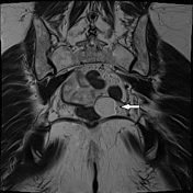
OvalWhite arrow: high signal oval mass posterior to the vaginaleft, coming close to thenear rectum in the lower side.
The mass has smooth margins, homogenous low signal on T1 and hyperintense on T2 even on the fat-sat sequence with homogeneous enhancement post-gadolinium. It shows hyperintensity of signal on DWI, without any diffusivity restriction on the ADC map. It has the appearance of fluid, no haemoglobin degradation products seen
Image Annotated image (Axial T2) ( update )

Image Annotated image (Axial T1 C+) ( update )
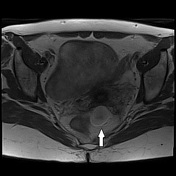
Image Annotated image (Axial T1) ( update )
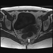
Image Annotated image (Sagittal T2) ( update )
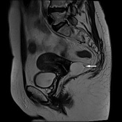
Image Annotated image (Coronal T2) ( update )
Updates to Case Attributes
GynaecologicalGynecological examination:
- nodular neoformation hard - smooth, posterior in the fornix
- speculum: epithelial portions: dystrophies, no atypical vaginal discharge
TV ultrasound:
- rounded hypoechoic mass with well-defined margin and 25 mm in diameter at the back of the cervix (myoma of the ligament?) non-liquid swelling free endopelvic. No ovarian abnormalities.
In the MRI report it was suggested to remove the mass because there were signs that were concerning for malignancy.
Histological findings: The neoformation shows a proliferation of elements sometimes fused in a pattern myxo-fibrosarcoma-like at the most distinct pleomorphic plurinucleates with MFH-like aspects. There are numerous lipoblasts often pleomorphic immersed in a myxoid stroma rich in capillary vessels. The mitotic index is high (> 3-4 myths xhpf), no necrosis sites are observed and the neoplasia reaches the margins of surgical excision. Immunocytochemistry proliferating elements express positive for CdK4 (clone EPR4513-32-7), CD 34 (clone QBEnd /10), MDM2 (clone IF2), S-100 (polyclonal) and Vimentina (clone V9) while negative for 1-A-2-N-EMA (clone E29), desmin (clone DE-R-11), cytokerite (clone AE1-AE3) and smooth muscle alpha-actin (clone 1A4). KI67 (clone 30-9) is expressed in 20% of proliferating elements.
-<p>Gynaecological examination:</p><ul>- +<p>Gynecological examination:</p><ul>