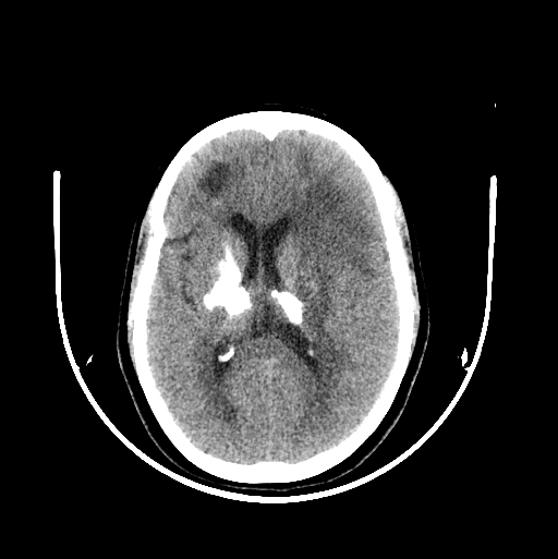
CT
Cystic lesions involving bilateral frontal deep white matter with surrounding oedema and areas of calcification.
Chunky areas of calcification seen in bilateral basal ganglia, thalami and dentate nuclei.