Parasternal long axis view
 Parasternal long axis view
 Parasternal long axis view
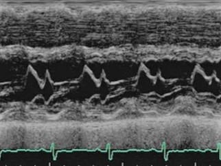
An M-mode echocardiogram from the parasternal long axis, with the m-mode pick directed through the mitral valve leaflets. One may appreciate the approximated mitral valve leaflets at the beginning of diastole (D) swing apart, with the cephalad and anterior excursion of the anterior leaflet contacting the septum at its peak excursion (E). As the pressure gradient between the left atrium and left ventricle begins to decline towards diastasis, the valves return toward their nuclear position (F). Their path is then reversed by atrial contraction (A), and thereafter trend toward one another until ventricular systole forces rapid coaptation.
Anatomy is labeled as RVOT (right ventricular outflow tract), IVS (interventricular septum), LV (left ventricle), and LVPW (left ventricular posterior wall).