
Presentation
Dysphagia for both solids and liquids with chest discomfort.
Patient Data
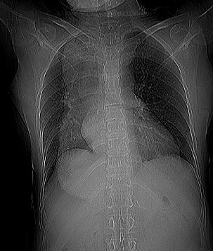
Large opacity overlapping the right mediastinum and hemidiaphragm of posterior location with absent gastric bubble, most likely due to dilated oesophagus filled with retained secretions and food.


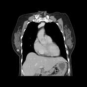
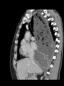
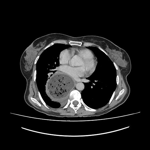
Grossely dilated oesophagus with moderately thickened wall, filled with fluid/food debris with smooth distal tapering at the gastro-oesophageal junction "bird beak sign" with no mass lesion. Collapsed normal stomach.
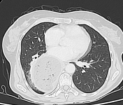
The lung window shows small cylindrical bonchiectasis of the right lower lobe with adjacent consolidation as well as small thin-walled lung base cyst.
No mediastinal or hilar lymphadenopathy seen.
Case Discussion
CT features of marked dilatation of the oesophagus, filled with fluid/food debris with smooth distal tapering at the gastro-oesophageal junction most consistent with achalasia.
CT has little role in directly assessing patients with achalasia but is useful in the assessment of the common complications and to identify any focal thickening which may indicate malignancy.


 Unable to process the form. Check for errors and try again.