
Presentation
Chest pain
Patient Data
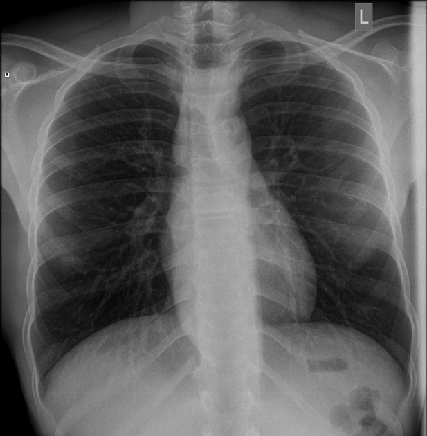
Frontal chest radiograph shows an undulating right paraspinal opacity that partly traverses the level of the diaphragm. Lucency projected over the mid trachea - concern for a dilated oesophagus, with the alternative being extramedullary haematopoiesis. An air-fluid level is not evident but may coincide with a vertebral endplate.
No consolidation, hilar lymphadenopathy or pleural effusion. Normal bones and soft tissues.
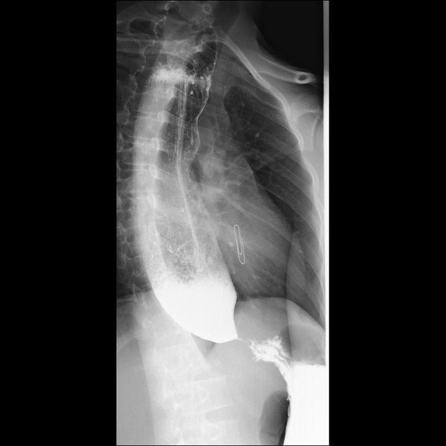
Contrast swallow showed a "rat's tail" constriction in the gastrooesophageal junction with marked upstream dilatation of the oesophagus and a proximal air-fluid level. Findings are typical of achalasia.
Case Discussion
The radiograph in this case is a good example of the importance of scrutiny of review areas in apparently normal radiographs. The retrocardiac and paraspinal regions often bear significant pathology and should be carefully evaluated.
Manometry is considered as the gold standard for the diagnosis of achalasia, and on its basis, three distinct subtypes are described 1. However, barium swallow studies may show features highly suggestive of this diagnosis in approximately 60% of cases 1, with the classic sign being tapering of the distal oesophagus (rat’s tail or bird’s beak sign).
Symptoms include dysphagia to both solids and liquids that characteristically develop at the same time 2. Chest pain, discomfort and eventual regurgitation complete the typical presentation.


 Unable to process the form. Check for errors and try again.