
Presentation
Left Achilles tendon region laceration a few hours before the presentation.
Patient Data
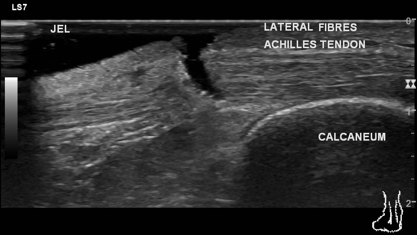
The lateral fibres of the Achilles tendon show a full-thickness defect approximately 25 mm away from the insertion site. There is no significant retraction. The central fibres of the tendon show a partial thickness defect involving superficial fibres. The medial most fibres of the tendon are intact. There is a defect in the overlying skin and subcutaneous tissue. The rest of the tendon shows normal echopattern.
There is no effusion in the retro-calcaneal bursa. The retrocalcaneal fat pad is normal.
Case Discussion
The case is an example of two useful concepts in the musculoskeletal ultrasound. One is the use of a gel pad. The defect is better characterised by a standoff jet pad as shown in the images. The other is the concept of a partial and full-thickness tear. The central fibres of the tendon show partial thickness tear involving superficial fibres. However, there is a full-thickness tear of the lateral most fibres.
Surgical repair of the tendon was done.


 Unable to process the form. Check for errors and try again.