
Presentation
First day newborn with dysmorphic features, short limbs and narrow chest.
Patient Data
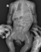
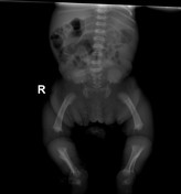

CHEST AND PELVIS X-RAYS:
Borderline cardiac size.
Increased left upper zone density probably due to enlarged thymus and rotation, less likely left upper zone consolidation clinical correlation is advised.
A progressive decrease in the interpedicular distance in the lumbar spine is seen, with widening of intervertebral disc spaces in this region.
Square shape iliac bones. (tombstone or mickey mouse ear) iliac wings
Short sacroiliac notches.
Flat acetabular roofs, with trident appearance of the acetabulum.
Upper and lower limb bone shortening is seen more proximally. (rhizomelic shortening)
Metaphyseal flaring with mesial bowing of lower limbs.
Long fibula, with the fibular head, is seen above the level of the tibial plateau.
Case Discussion
First day newborn on physical examination evaluation short limb, narrow chest and dysmorphic features were appreciated. Further evaluation of the skeletal system and other abnormalities was requested for suspected dwarfism. Unfortunately, lateral views were not provided. Anteroposterior narrowing of the ribs was appreciated on physical examination which is explained by non-visualization of the right heart border at the right paraspinal region due to displacement of the cardiac shadow to the left. Sharing radiological findings of Rickets, these imaging findings in newborns are classical features of achondroplasia, the most common type of bone dysplasia compatible with life.


 Unable to process the form. Check for errors and try again.