
Presentation
Shoulder trauma following a road traffic accident.
Patient Data
Age: 65 years
Gender: Female
From the case:
Acromioclavicular joint dislocation


Show annotations
Download
Info
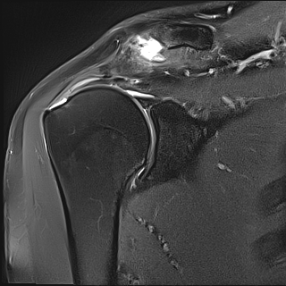
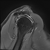
acromioclavicular dislocation with probable tear of the coracoclavicular ligament
bony flecks avulsion from the distal aspect of the clavicle
full-thickness tear of the supraspinatus tendon
mild articular surface tear of the footprint of the infraspinatus tendon
-
mild soft tissue oedema and joint effusion
From the case:
Acromioclavicular joint dislocation
Download
Info

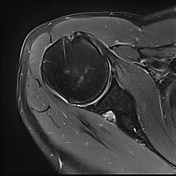
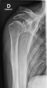
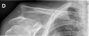
Dislocation of the right acromioclavicular joint with an elevation of the clavicle above the superior border of the acromion (the coracoclavicular distance is still less than twice normal)
From the case:
Acromioclavicular joint dislocation

Download
Info
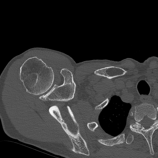
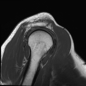
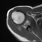
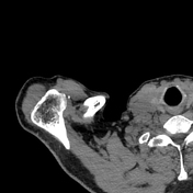
The small bone fragments are best seen on CT.
Case Discussion
Features of an acromioclavicular joint injury type III of the Rockwood classification and a full-thickness rotator cuff tear.


 Unable to process the form. Check for errors and try again.