
Presentation
Lower abdominal pain, right more than left. Raised white cell count and CRP.
Patient Data


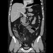
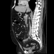
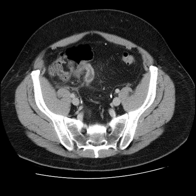
The appendix is located medial to the caecum and is abnormally dilated, demonstrating prominent mural enhancement. There is fat stranding surrounding the appendix, along with thickening of the pelvic parietal peritoneum on the right.
Pathology report:
Macroscopic: the appendix serosa is irregular. Parts of the appendix are covered by a fibrinopurulent exudate. The mucosa is normal. No tumour or faecolith.
Microscopic: sections of the appendix demonstrate numerous neutrophils in the lumen and the wall. No tumour.
Pathology diagnosis: acute appendicitis.
Case Discussion
The imaging features are consistent with acute appendicitis without evidence of perforation.


 Unable to process the form. Check for errors and try again.