
Presentation
Four days history of fever, nausea, vomiting and truncal ataxia
Patient Data
Age: 7 years
Gender: Male
From the case:
Acute cerebellitis


Download
Info

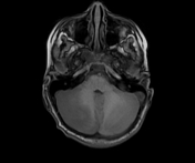
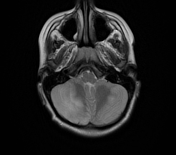
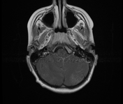
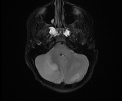
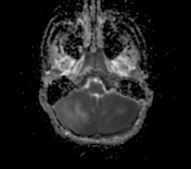
The MRI sequences demonstrate:
- swelling of both cerebellar hemispheres with ill-defined areas of low signal on T1WI, high signal on T2WI/FLAIR involving both grey and white matter (cerebellar oedema) with regions of restricted diffusion as well as increased diffusivity on ADC map. The multivoxel MR spectroscopy shows low NAA/Cr and NAA/Cho ratios
- mild leptomeningeal enhancement is noted along the cerebellar hemispheres mainly on the right
- a mass effect on the 4th ventricle with tonsillar herniation. Mild upward cerebellar displacement with a mass effect on the brainstem and quadrigeminal cistern
- dilated third and lateral ventricles in keeping with obstructive hydrocephalus
Case Discussion
MRI features are most consistent with an acute cerebellitis.
Additional contributors: A. Ramdani, MD / ZE. Boudiaf, MD


 Unable to process the form. Check for errors and try again.