
Acute cerebral infarction due to MCA stenosis caused by vasculitis
Presentation
Headache, right-sided paralysis, and sensory disturbances.
Patient Data


The following lesions were noted on MRI:
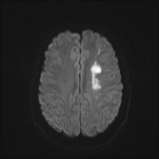
scattered acute cerebral infarctions in the left hemisphere (frontal, temporal, parietal regions, putamen, and centrum semiovale), with true diffusion restriction
MRA shows severe stenosis of the proximal M1 segment of the left middle cerebral artery
on vessel wall MRI with pre- and post-contrast T1FS SPACE sequences, concentric thickening of the proximal M1 segment of the left middle cerebral artery is noted, with strong contrast enhancement after injection
Case Discussion
The patient shows no signs of systemic infection clinically. Blood tests indicate elevated CRP and D-Dimer levels, no anemia, and an erythrocyte sedimentation rate within normal limits. Cerebrospinal fluid tests are normal.
The MRI findings and clinical symptoms are consistent with acute cerebral infarction due to middle cerebral artery stenosis caused by vasculitis (possible central nervous system vasculitis).
Vessel wall MRI helps to detect various vascular lesions causing stenosis or occlusion of intracranial arteries, such as:
intracranial atherosclerotic disease (ICAD): eccentric wall thickening, usually with heterogeneous enhancement
vasculitis: concentric wall thickening with homogeneous enhancement
reversible cerebral vasoconstriction syndrome (RCVS): vessel wall thickening with no enhancement or minimal enhancement
arterial dissection: intimal flap seen on T2-weighted images, with high signal blood within the arterial wall on T1-weighted images
moyamoya disease: stenosis of the internal carotid artery without enhancement


 Unable to process the form. Check for errors and try again.