
Acute ICA ischaemic penumbra due to high-grade CCA stenosis (CT perfusion)
Presentation
Acute onset slurred speech, facial droop and left-sided hemiparesis. Reduced GCS. Previous CVA (right ACA) 6 months earlier.
Patient Data
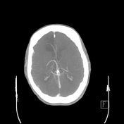
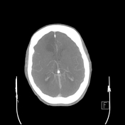
Non contrast CT
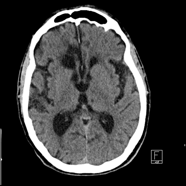
Hypodense gliosis in the right ACA territory in keeping with prior infarct. Similar post-ischaemic gliosis in the left temporal lobe. No new area of grey-white matter differentiation loss. No insular ribbon sign. No dense vessel sign evident on the thin data set. No haemorrhage, surface collection, mass effect or midline shift. Ex vacuo dilatation of the right lateral ventricle. Prominent CSF in the right CP angle with mild mass effect on the right cerebellar hemisphere is an arachnoid cyst which is stable.
CT angiogram
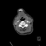

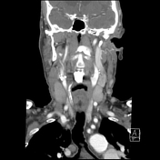
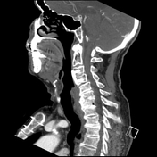
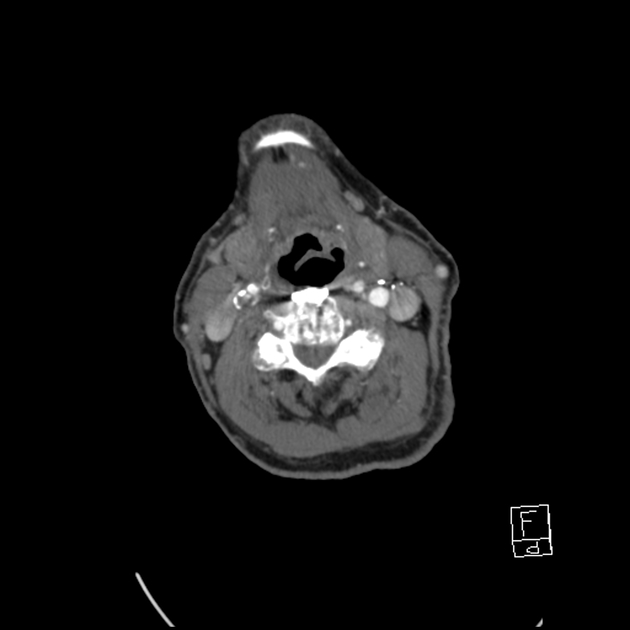
No intracranial vascular occlusion identified. High-grade stenosis due to calcified and noncalcified plaque of the dominant right V4. The nondominant left vertebral artery terminates as a PICA. Heavily calcified right vertebral artery, bilateral cavernous ICAs and right terminal ICA. The right common carotid artery bifurcation demonstrates irregular calcified and noncalcified plaque which extends into the proximal ICA causing a high-grade ICA stenosis. Surgical clips anterior to the left carotid sheath. Dural venous sinuses enhance normally.
CT perfusion
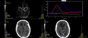
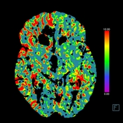
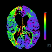

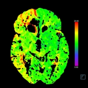
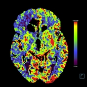

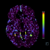
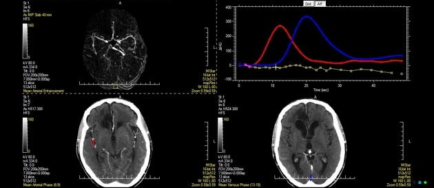
The old right ACA territory infarct demonstrates markedly decreased CBV and CBF as expected. Similar changes in the left temporal pole.
Decreased MTT, Tmax and TTD in the right cerebral hemisphere affect the ACA and MCA territories and spares the occipital lobe (PCA territory). No corresponding CBF or CBV abnormality on the right to suggest core infarct.
Case Discussion
- no haemorrhage or signs of acute infarction. No large vessel occlusion
- severe complex right CCA bifurcation/proximal ICA atheromatous plaque causing high-grade stenosis and near-complete occlusion
- single right vertebral artery supplies the basilar artery, with a high-grade atheromatous stenosis of V4
- perfusion colour maps demonstrate increased time parameters reflecting penumbra in the right cerebral hemisphere due to the high-grade stenosis of the ICA. These changes could also be secondary to seizure but there was no indication of this in the clinical presentation. No core infarct identified
- old right ACA infarction
The patient was not suitable for IV thrombolysis.


 Unable to process the form. Check for errors and try again.