
Presentation
Backache for 1 month.
Patient Data


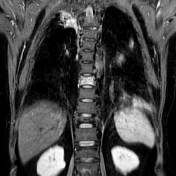
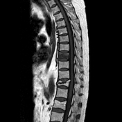
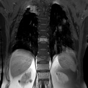
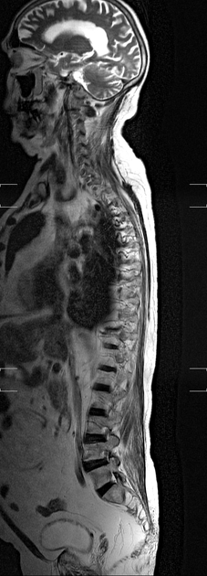
There is reduction in height of the T8 vertebral body with mild biconcave appearance. Altered marrow signal noted within which appears T1/T2 hypointense, STIR hyperintense with mild post-contrast enhancement. No associated abnormally enhancing soft tissue noted.
-
Compression fracture of the T11 vertebral body with retropulsion of the posterior fractured segment, effacing the thecal sac and indenting the cord without abnormal cord signal. No post-contrast enhancement noted within the vertebra.
Case Discussion
In this patient with osteoporosis, with typical biconcave appearance of the vertebra, the altered marrow changes are consistent with acute osteoporotic fracture.
Pathological fracture due to malignancy is less likely due to:
No expansion of the vertebral body.
No associated soft tissue.
No involvement of the posterior elements.


 Unable to process the form. Check for errors and try again.