
Presentation
History of seizures and visual disturbance.
Patient Data
Age: Child
Gender: Male


Download
Info
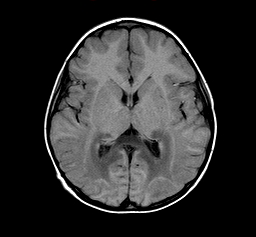
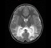
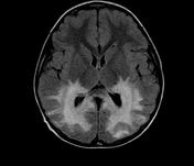
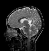
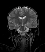
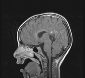
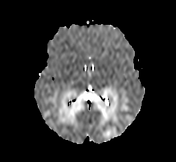
There is evidence of bilateral symmetrical confluent low Tl signal and high T2 signal involving peritrigonal white matter, optic radiation as well as splenium and extending to involve the corticospinal tracts. The subcortical U-fibre white matter is spared. The "leading edge" also demonstrates enhancement after Gadolinium injection. Additionally, there is bilateral symmetrical auditory pathways involvement at the level of brain stem (pons) with enhancement after contrast injection.
Case Discussion
These MRI features are typical for X-linked adrenoleukodystrophy and the diagnosis of this case was confirmed.


 Unable to process the form. Check for errors and try again.