
Presentation
History of chronic sinusitis in an asthmatic patient with a right nasosinusal mass on ENT exam.
Patient Data


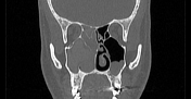
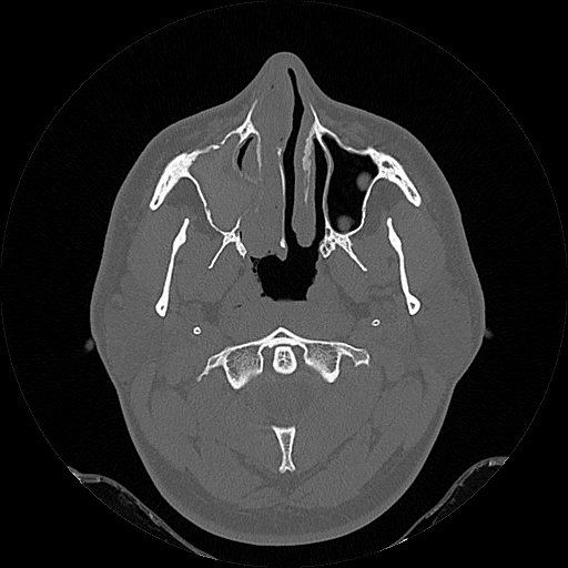
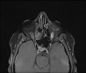
Soft tissue filling the right sinuses (maxillary, sphenoid, frontal, and ethmoid cells), isodense to the muscles with central areas mildly hyperdense. There is a remodelling and thinning of the bone sinus wall with the expansion of the maxillary ostium into the ipsilateral nasal fossa.
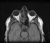
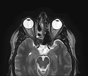
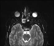
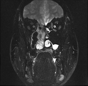
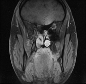
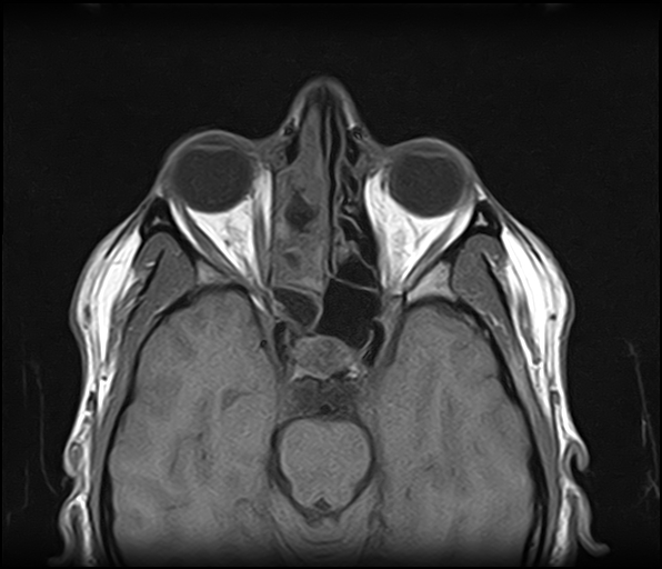
The MRI sequences demonstrate a hypointense signal on T1WI, markedly hypointense signal on T2WI mainly of the maxillary antrum and sphenoid sinus (due to the high concentration of various metals by the fungal organisms as well as the high protein content) with peripheral inflamed mucosal thickness of intermediate signal on T1WI, high signal on T2WI, enhanced on post-contrast sequences. The right ethmoid cells as well as the ipsilateral nasal fossa show also areas of very low signal on T2WI. Moderate peripheral mucosal thickening of the left maxillary antrum.
Case Discussion
CT and MRI features are highly suggestive of an allergic fungal sinusitis
The allergic fungal sinusitis is considered as the most common fungal sinusitis. The treatment includes local excision with steroid therapy. The fungal therapy is used in some cases.
Additional contributor: Dr. R Bouguelaa, MD


 Unable to process the form. Check for errors and try again.