
Presentation
Memory probelms.
Patient Data


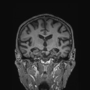
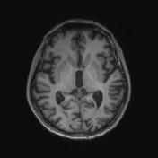
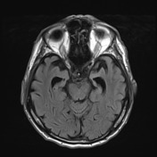
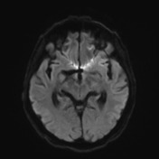
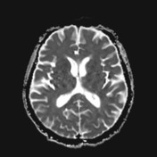
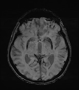
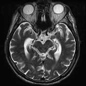
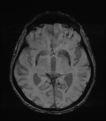
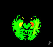
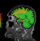
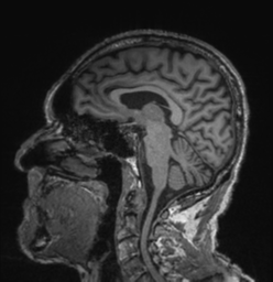
There is mild generalised cerebral volume loss without particular lobar predominance. Bilateral hippocampal atrophy is demonstrated, more than the degree of the cerebral volume loss elsewhere, more so on the left side (ERICA score = 2 on the right and = 3 on the left). There is mild chronic small vessel ischaemic change. No hydrocephalus. No space-occupying lesion. No abnormal susceptibility to suggest cerebral amyloid angiopathy. No abnormal diffusion restriction. The remainder of the brain is unremarkable.
Conclusion:
In this clinical context, findings are compatible with Alzheimer's disease or limbic-predominant age-related TDP-43 encephalopathy (LATE).
Case Discussion
Unfortunately at this time, there are no additional tests (e.g. amyloid PET or CSF) available to help distinguish between the two entities.


 Unable to process the form. Check for errors and try again.