
Presentation
Memory difficulties.
Patient Data
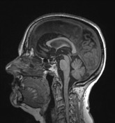

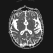
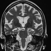
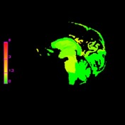
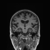

The hippocampi are small bilaterally reduced in volume, with associated widening of the choroidal fissures and temporal horns. There is additional less marked volume loss within the temporal lobes and cingulate gyrus. Note: Sagittal segmented volume loss (z-score deviation from age-matched healthy population) were created using prototype software not available for commercial or clinical use.
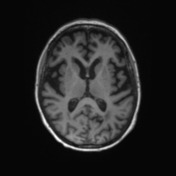
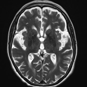
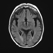
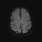
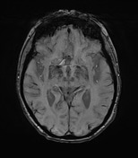
No intra extra-axial mass, collection or focal abnormality. No evidence of cerebral amyloid angiopathy. No abnormal restricted diffusion. A small amount of patchy white matter T2 signal hyperintensity is consistent with chronic small vessel ischaemic change. Ventricles are commensurate with the degree of cerebral volume loss.
Case Discussion
The pattern of hippocampal volume loss, some parietal loss and memory loss in a 75 year old is very suggestive of Alzheimer's disease.


 Unable to process the form. Check for errors and try again.