
Presentation
4 years history of short-term memory loss
Patient Data
Age: 60 years
Gender: Male


Download
Info
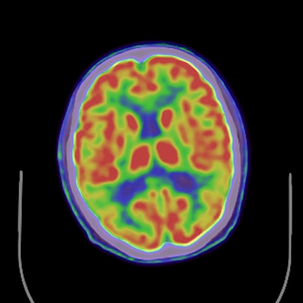
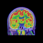
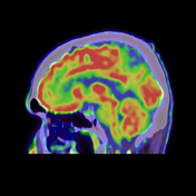
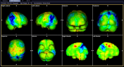
- There is significant hypoperfusion involving the temporal parietal region bilaterally. This is largely symmetrical in appearance.
- Note is also made of hypoperfusion involving the medial temporal lobes, posterior cingulate gyrus and precuneus.
- Minor patchy areas of hypoperfusion identified in the frontal lobes bilaterally.
- No definite hypoperfusion appreciated elsewhere.
- Normally preserved perfusion of the primary sensory motor cortex, basal ganglia, thalamus and cerebellum.
Case Discussion
The pattern of cerebral hypoperfusion predominantly involving the temporoparietal lobes bilaterally is typical of Alzheimer's disease. This is further substantiated by the patient's clinical history and was supported by his clinician.


 Unable to process the form. Check for errors and try again.