
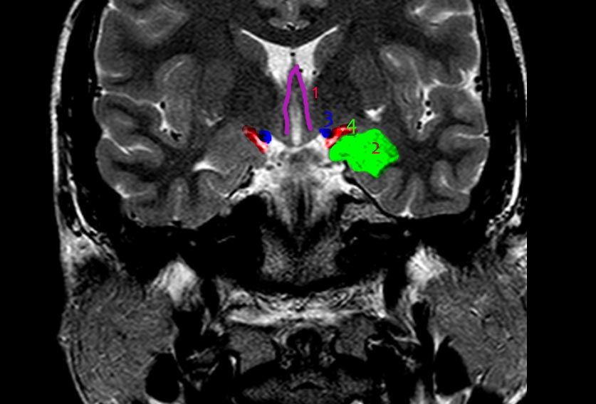
The anatomy of the amygdala.
The endorhinal sulcus is the anatomical landmark for the anterior extension of the amygdala identified by the red arcuate line (4). The amygdala: the green structure (2). The two blue dots represent the optic tracts (3). The vertical purple lines represent the anterior limbs of the fornix diving through the hypothalamus (1).
Illustrations and annotations created by Azza Elgendy
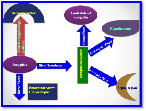
The main output (efferent) fibres of the amygdala:
Ventral amygdalofugal pathway: longer pathway dives through the septal region, brain stem, thalamus, hypothalamus, and finally into the sensory cortex.
-
Stria terminals (very similar to the fornix in relation to the anterior commissure); it is divided into three parts in relation to the anterior commissure:
pre-commissure fibres
commissure fibres
post-commissure fibres
Illustration created by Azza Elgendy
Case Discussion
Amygdala anatomy is illustrated and explained.


 Unable to process the form. Check for errors and try again.