
Amyloid arthropathy and myopathy, mimicking osteomyelitis with intramuscular abscess
Presentation
Underlying end-stage renal failure, history of renal transplant, presented with fever, chills, poor oral intake. CRP was persistently elevated.
Patient Data
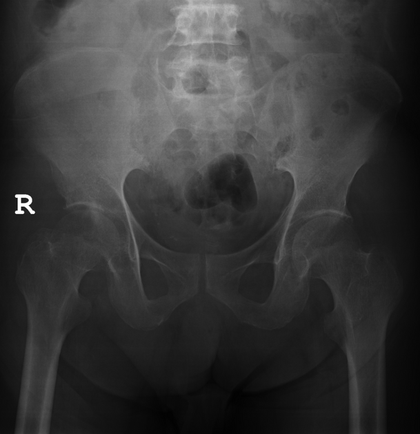
Subtle bony erosion of the right posterior acetabulum.


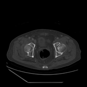
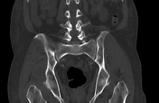

Bony erosions of the right ischium (posterior acetabulum) with a rim-enhancing hypodense lesion involving the right gluteus minimus muscle. Rim-enhancing hypodense lesions were also noted involving bilateral gluteus medius muscles (just underneath iliotibial bands), more prominent on the left. Smaller bony erosions at bilateral proximal femora.
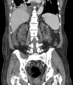
Additional findings include polycystic kidneys, an atrophied transplanted kidney at the right iliac fossa, free intraperitoneal fluid, cholelithiasis and bilateral pleural effusions.
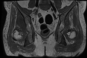

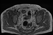
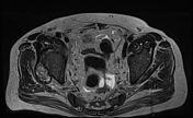
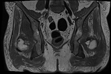
Deposition of soft tissue lesions with abnormal low T1 and T2 signals (compared to bone) at the following locations:
bilateral proximal femora, associated with subchondral erosions
between left gluteus maximus and gluteus medius muscles
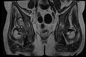
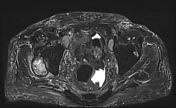
Deposition of soft tissue lesions with abnormal low T1 and heterogenous high T2 signals (compared to bone) at the following locations:
right ischium (posterior acetabulum) with bone erosion and indentation onto right gluteus minimus muscle, associated with muscle oedema
left ischium with minimal bony erosion
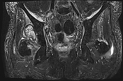
bilateral gluteus medius muscles located just underneath iliotibial bands, more prominent on the left
bilateral iliopsoas muscles, subcentimetre
Between bilateral gluteus maximus and gluteus medius muscles, subcentimetre.
These lesions mentioned above are T2 hyperintense (compared to skeletal muscles) on fat saturation sequence.
Minimal marrow oedema adjacent to the aforementioned bony erosions.
Synovial thickening of bilateral hip joints, worse on the right. No significant hip joint effusion.
Sacroiliac joints and symphysis pubis are preserved.
Minimal ascites in the pelvis. Minimal subcutaneous oedema near the coccyx.
Summary:
With the given clinical information, MRI findings likely represent amyloid arthropathy and myopathy. Suggest correlation with HPE results.
Case Discussion
Ultrasound-guided biopsy of the right ischial/posterior acetabular lesion was performed.
HISTOPATHOLOGY REPORT :
Macroscopy:
Appearance: Longitudinal strips of brownish tissue
Number of strips: 3
Measurement: 3 - 19 mm in length
Submitted entirely in 1 block.
Microscopy:
Levels show three strips of cartilaginous tissue with adjacent fibrous and adipose tissue. There are foci of amorphous eosinophilic material deposition seen. There is no evidence of malignancy.
Special stain: Congo Red shows apple-green birefringence under polarised light.
Interpretation: Consistent with amyloid arthropathy.
Case courtesy of Dr Noorfizura Binti Ahmad, Consultant Radiologist, Sarawak General Hospital, Malaysia.


 Unable to process the form. Check for errors and try again.