
Presentation
Back and hip pain.
Patient Data
Age: 55 years
Gender: Male
From the case:
Ankylosing spondylitis

Download
Info
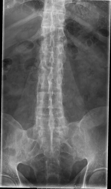
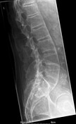
Lumbar marginal syndesmophytes giving bamboo spine. Calcification of the interspinous ligament is seen as well (dagger sign).
Bony fusion of bilateral sacroiliac joints is noted, suggestive of chronic sacroiliitis.
From the case:
Ankylosing spondylitis


Download
Info
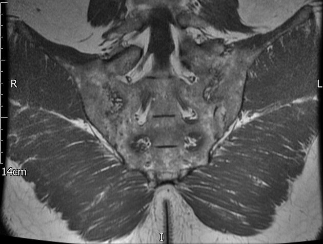
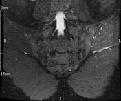
MRI of the sacroiliac joints shows bilateral bony fusion and subchondral fatty changes in keeping with chronic changes.
There are no evident signs of active spondyloarthropathy and no subchondral foci of bone marrow oedema.
Case Discussion
A Known case of ankylosing spondylitis. MRI is useful to check disease activity in the form of bone marrow oedema and to plan management accordingly.


 Unable to process the form. Check for errors and try again.