
Presentation
The patient presented with long standing back pain.
Patient Data
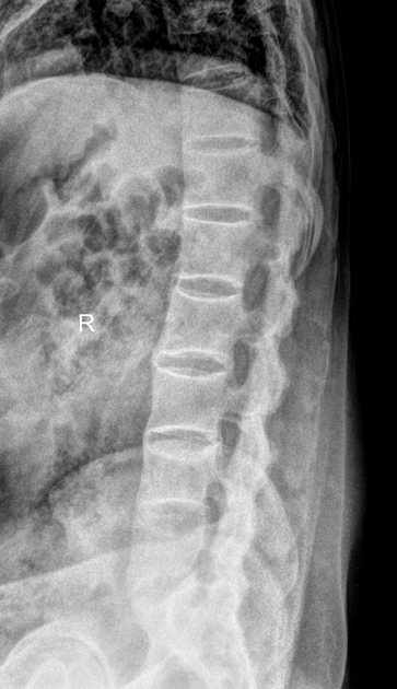
There is flowing paravertebral ossification on both lateral and AP projections with preserved disc space giving the appearance of a bamboo spine. Ossification of the interspinous and supraspinous ligaments giving the appearance of a dagger spine is also noted. Complete ankylosis of the bilateral sacroiliac joints is also visualised as well as ankylosis of the bilateral facet joints.
There is also vertebral body squaring with loss of anterior concavity of the anterior vertebral border.
There is discontinuity of the anterior and posterior bridging syndesmophytes at the L5/S1 level suspicious for chalk stick fracture.
Case Discussion
Ankylosing spondylitis (AS) is seronegative spondyloarthropathy which results in fusion (ankylosis) of the spine and sacroiliac (SI) joints. Patients are rheumatoid factor (RF) negative, hence seronegative.
The axial skeleton is predominantly affected with sacroiliitis as the earliest manifestation, although in ~20% of cases, the peripheral joints are also involved. Erosion and ankylosis are the hallmarks of the disease.
MRI is more sensitive than conventional radiography in detecting early inflammatory changes such as synovitis, capsulitis and enthesitis.
In our cases, the disease is in advanced stage evidenced by complete ankylosis of the bilateral sacroiliac joints.


 Unable to process the form. Check for errors and try again.