
Presentation
The patient presented with subacute headache episodes that have not responded to medical therapy.
Patient Data


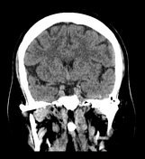
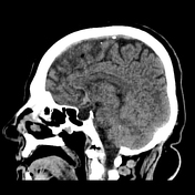
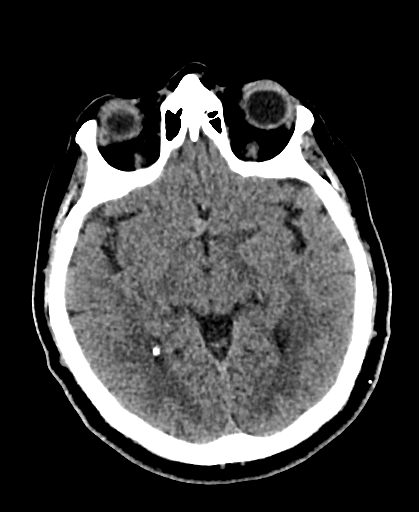
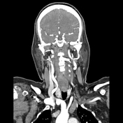
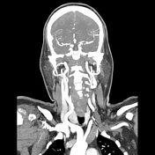
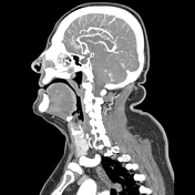
A hyperattenuating nodule is observed in the anterior arterial territory, located near the floor of the third ventricle.
No evidence of intracranial haemorrhage.
There was a suspicion of an aneurysm based on imaging findings, prompting further investigations to confirm the diagnosis and assess its implications.

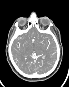

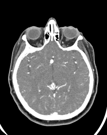
A saccular dilation is seen at the junction where the anterior cerebral arteries converge in keeping with anterior communicating artery (ACom) aneurysm.
The imaging shows a small calibre of the left A1 segment of the anterior cerebral artery.
Case Discussion
After reviewing the non-contrasted study, there was a high suspicion of an anterior communicating artery (ACom) aneurysm. Subsequently, contrast-enhanced images were obtained to further evaluate and confirm the presence, size, and characteristics of the aneurysm.
Aneurysms pose a risk of rupture and are typically evaluated and managed through imaging studies such as CT or MRI, followed by interventions like endovascular coiling or surgical clipping based on size and clinical considerations.


 Unable to process the form. Check for errors and try again.