
Presentation
Right side nasal cavity obstruction and nasal speech.
Patient Data
Age: 40 years
Gender: Male
From the case:
Antrochoanal polyp


Download
Info
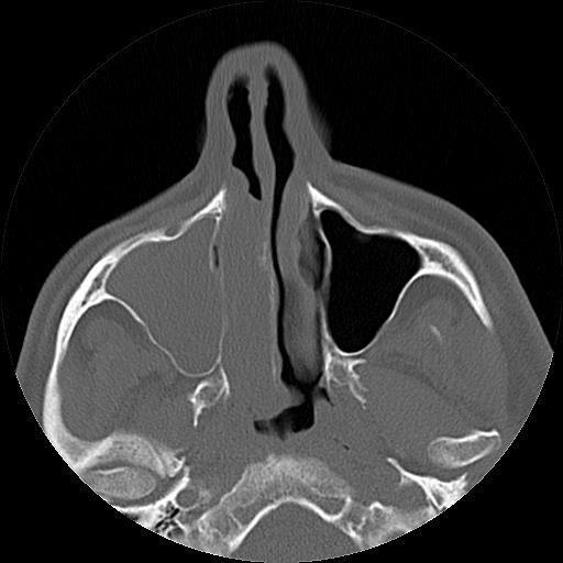
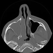
Large mass-like lesion within the right maxillary sinus opacified and expanded the sinus antrum with growth within the sinus ostium, expanded the ostium and extended within the right side nasal cavity middle and inferior meatus, and bulged within the ipsilateral nasopharynx is seen.
Case Discussion
The case illustrates typical non-contrast CT scan features of an antrochoanal polyp. One of the complications of the antrochoanal polyp is papillary endothelial hyperplasia which can lead to hemorrhage. The polyp has usually a narrow pedicle but rarely can be seen on CT scan images. The treatment of choice is complete surgical resection of the pedicle.


 Unable to process the form. Check for errors and try again.