
Presentation
Right lower quadrant pain. Appendix not located on ultrasound.
Patient Data


Small hiatal hernia.
Esophageal, left gastric, and retrogastric varices.
Small retrocrural nodes, visible on the previous study 7 years prior (not shown).
Nonobstructing portal confluence thrombus, extending into the portal vein, probably longstanding.
Enlarged lymph node between the liver and pancreatic body, unchanged.
The gallbladder has been removed.
Dilated central intrahepatic bile ducts, not dilated on the previous study.
The spleen is enlarged, has grown since the previous study.
Right renal cortical cyst 2.7 cm in diameter.
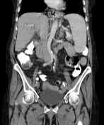
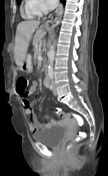
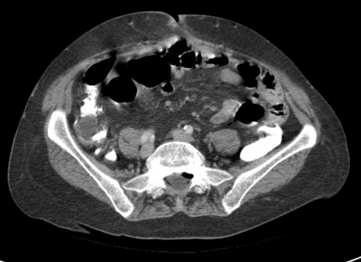
The vermiform appendix is dilated up to 19 mm by fluid content, similar to the previous study.
Tiny amount of free fluid posterior to the uterus.
Two small epigastric fat-containing hernias.
Summary:
- Portal vein thrombosis, with portosystemic collaterals and an enlarged spleen, suggest portal hypertension.
- Appendiceal mucocele.


 Unable to process the form. Check for errors and try again.