
Presentation
Work up for abdominal pain.
Patient Data
Age: 70 years
Gender: Male
From the case:
Appendicolith

Show annotations
Download
Info

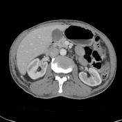
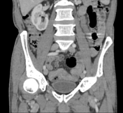
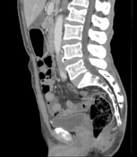
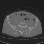
Few calcifc foci are noted in the lumen of the appendix however, its luminal diameter is within normal limits.
No surrounding stranding is seen.
No free fluid is noted.
The rest of the viscera appear unremarkable.
Case Discussion
CT findings most likely suggest appendicoliths without CT evidence of appendicitis as described above.
Co-contributor: Dr, Anwar-ul-Haq Zadran.


 Unable to process the form. Check for errors and try again.