
Presentation
Fever; frequent fall while trying to stand up from sitting position; weakness of bilateral lower limbs.
Patient Data

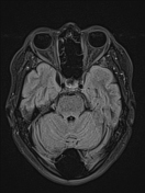
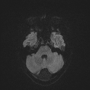

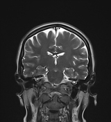
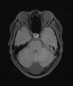
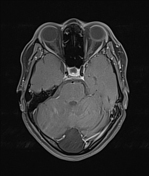
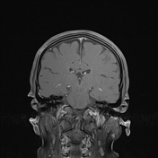
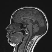
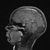
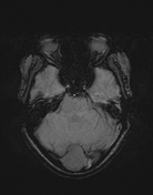

A well-defined, oval-shaped, T1 hypointense/ T2 hyperintense cystic lesion which suppresses on FLAIR is noted in the posterior cerebellar region. DWI/ ADC shows no diffusion restriction. SWI shows no blooming artefact. Post-contrast study shows no enhancement. Anteriorly it slightly compresses the cerebellar vermis and the bilateral cerebellar hemispheres on each side. Posteriorly it causesslight scalloping of the adjacent occipital bone. However, the ventricles are normal and no definite communication with the 4th ventricle is seen.
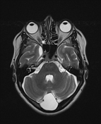
CSF space in the right medial temporal region is prominent.
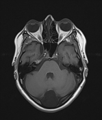
There is mucosal thickening with T2/ FLAIR high signal intensity noted in the right maxillary sinus which shows enhancement in T1 post-contrast study suggesting sinusitis.
Case Discussion
A well-defined, oval-shaped, non-enhancing, cystic lesion in the posterior cerebellar region with scalloping of adjacent occipital bone with no obvious communication seen with the 4th ventricle and no associated hydrocephalus suggesting arachnoid cyst.


 Unable to process the form. Check for errors and try again.