
Presentation
A child presented with twitching of the left cheek, angulation of the mouth with excessive saliva and no history of trauma.
Patient Data


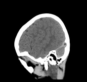
An incidentally noted, sharply circumscribed, oval filling defect within a straight dural venous sinus isodense relative to CSF density protrusion into its confluence /beginning with small pedicle, measuring about 6 mm in diameter.
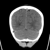
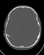
Another similar one is noted at the junction of the right transverse dural venous sinus, measuring about 5 mm typical of arachnoid granulations.
Conclusion:
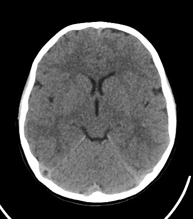
Apart from incidentally noted typical arachnoid granulations (normal variant). Otherwise an unremarkable enhanced CT brain study.
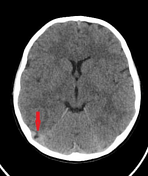
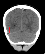
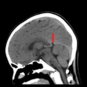
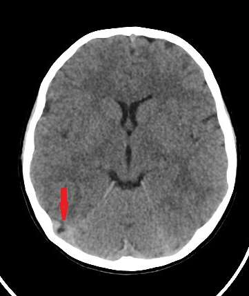
Red arrows on axial and coronal images pointing to the one in the right transverse sinus, on the sagittal image pointing to the other in the straight sinus.
Case Discussion
This is a good example of an incidentally noted typical arachnoid granulation. Typically, they range from 2 to 8 mm and occasionally can exceed 1 cm in diameter.
An arachnoid granulation typically appears on Contrast-enhanced CT (our case non-enhanced CT) as a round or oval filling defect within a dural venous sinus.
Arachnoid granulations are most commonly located within the transverse sinuses, superior sagittal sinus, and parasagittal venous lacunae.


 Unable to process the form. Check for errors and try again.