Presentation
Pre-employment screening chest radiograph.
Patient Data

Frontal chest radiograph shows prominent shadows of the pulmonary trunk, main branches, and aortic arch.
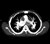

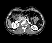

All the visualized arteries, including the major arteries and their branches, appear tortuous. However, they are patent and opacified with contrast, suggesting features of connective tissue disease, including arterial tortuosity syndrome.
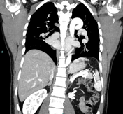
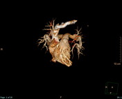
Coronal images show an aortic elongation sign; elongation of the aorta leading to prominent aortic knuckle.
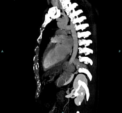
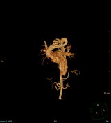
Sagittal images show a cluster of vessels sign; tortuosity of origins of aortic arch branches 2.
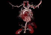
3D images show a tortuous aortic arch and its branches, subclavian artery, celiac trunk, splenic artery, main renal arteries, and left main pulmonary artery.
Case Discussion
A pre-employment screening chest radiograph of an otherwise healthy middle-aged male showed prominent shadows of the pulmonary trunk, main branches, and aortic arch.
Further work-up by CT with contrast showed that all the visualized arteries, including the major arteries and their branches, appear tortuous, without evidence of filling defects or thrombosis.
Physical and laboratory tests of the mentioned patient were unremarkable, however, regular follow up tests were recommended.
Features are suggestive of connective tissue disease including arterial tortuosity syndrome, which is an autosomal recessive connective tissue disorder characterized by:
extreme arterial tortuosity: this includes twisting and elongation of arteries, which can lead to complications such as aneurysms and dissections 1
pulmonary arterial stenosis: severe narrowing of the pulmonary arteries, causing right ventricular hypertension and dysfunction 1
systemic manifestations: these can include features like bifid uvula, skin and muscular changes, and joint laxity 2
Regular follow-up with MRA or CTA is advised to monitor the progression of arterial tortuosity and detect any new aneurysms or dissections early.
Clinical follow-up includes continuous monitoring of cardiovascular health and timely surgical planning are crucial for managing ATS effectively 1.


 Unable to process the form. Check for errors and try again.