
Presentation
History of about 20 years asbestos exposure.
Patient Data
Age: 75 years
Gender: Male
From the case:
Asbestos pleural placques


Download
Info

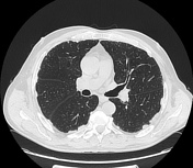
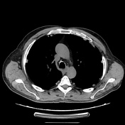
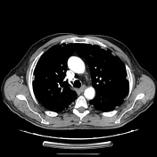
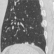
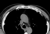
CT shows the presence of bilateral pleural plaques, few of them calcified, and subpleural reticular bands.
Aspecific lymph nodes are visible in the mediastinum.
The ascending aorta has a diameter at the maximum values of the standard (mm 40).
Cohesist the presence of multiple calcifications of the head and tail of the pancreas.
Case Discussion
The presence of pleural plaques represents the typical appearances of those subjected to long-term exposure and inhalation of asbestos fibres.
Traction bronchiectasis are other characteristic signs of this disease.


 Unable to process the form. Check for errors and try again.