
Presentation
Chronic cough and shortness of breath.
Patient Data


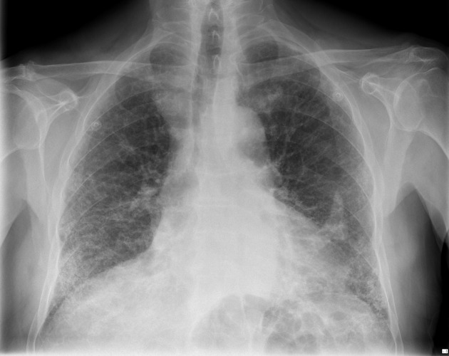
Bilateral reticular opacity, worse in the bases, with reduced lung volume in keeping with previously seen interstitial fibrosis, which appears to have progressed from the previous chest x-ray. Left sided calcified pleural plaques. No new areas of focal consolidation. No pleural effusion.


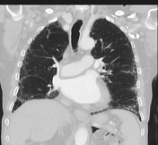
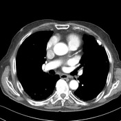
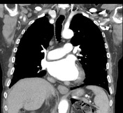
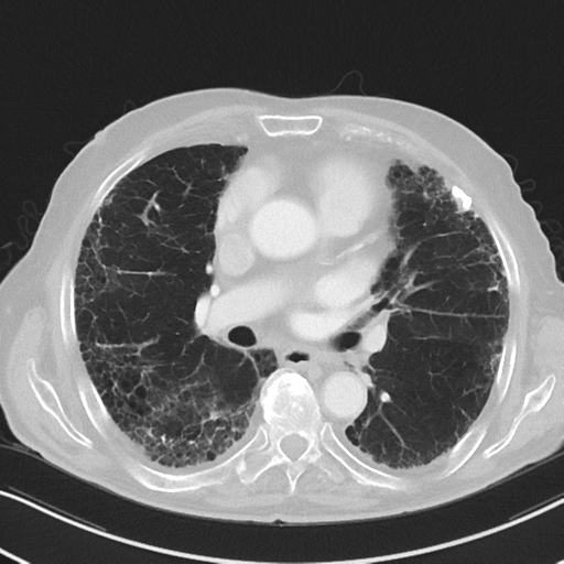
UIP pattern characterized by subreticular opacity, honeycombing and traction bronchiectasis worse in the bases has mildly progressed. No suspicious pulmonary nodule or mass. No focal consolidation or lobar collapse. Calcified pleural plaques are again seen. No pleural effusion. Tracheal and proximal bronchi are within normal limits. No enlarged thoracic lymph nodes.
Case Discussion
UIP pattern along with calcified pleural plaques is highly suggestive for asbestosis, and this patient had the correlative history of asbestos exposure.


 Unable to process the form. Check for errors and try again.