
Presentation
Weakness in bilateral upper limbs
Patient Data


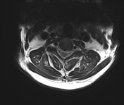
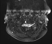
Partial fusion of anterior arch of atlas with clivus. Posterior arch of atlas is not visualised separately and is likely fused with occiput.
Atlantoaxial subluxation is seen with atlantodental interval being approx 9 mm.
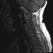
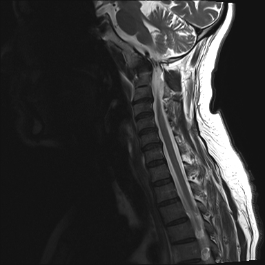
Basilar invagination is also seen with superior migration of dens of C2, projecting above foramen of magnum causing severe compression of cervicomedullary junction and upper cervical cord.
Hyperintensities are seen in cervicomedullary junction and upper cervical cord at C2 level.
Erosion/cystic change seen in dens of C2 with tiny calcifications at anterior aspect of dens and heterogeneous soft tissue in atlantodental interval - likely degenerative changes. Possibility of inflammatory arthritis, example rheumatoid appears less likely.
C2-C3 block vertebra seen.
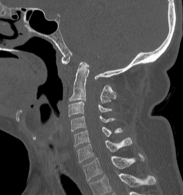
CT confirming the above findings on MRI.
Case Discussion
Overall above findings are suggestive of atlantooccipital assimilation with atlantoaxial subluxation, basilar invagination causing compressive myelopathy at cervicomedullary junction and upper cervical cord.


 Unable to process the form. Check for errors and try again.