
Presentation
History of left undescended testis post second stage Fowler Stephens.
Patient Data

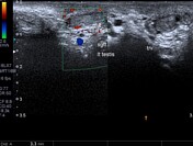

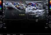
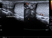
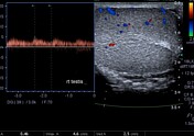
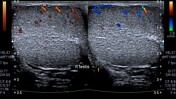
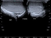
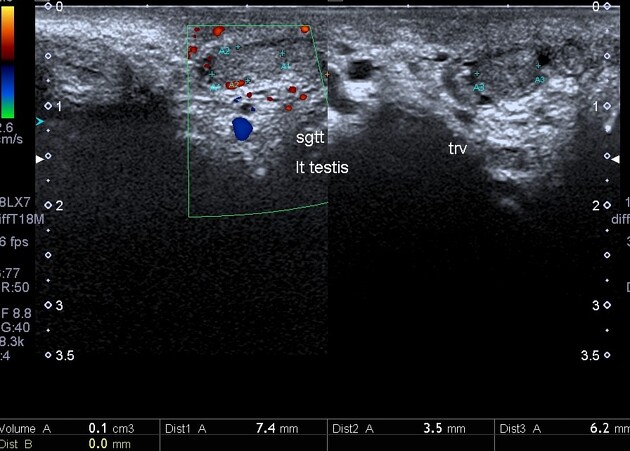
Both testicles are within the scrotal sac. There is unilateral left-sided testicular atrophy measuring 0.1 ml in volume with subtly reduced colour flow signals on colour/ power Doppler imaging. The right counterpart testis is well encapsulated and is compensatorily larger measuring 17 ml in volume albeit with unremarkable parenchymal echo/vascular pattern. Serpiginous vascular structures (varices) are noted bilaterally along the pampiniform plexi.
Case Discussion
Atrophic left testis post 2nd stage fowler stephens with compensatory hypertrophy of the right testicular counterpart and attendant Grade I bilateral scrotal venous varicoceles. No hydroceles, masses/calculi or inguinal-scrotal hernias seen.


 Unable to process the form. Check for errors and try again.