
Presentation
No trauma sustained. Sudden shortness of breath, ? pneumothorax.
Patient Data
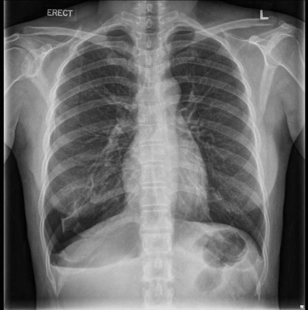
There is a right basal pneumothorax seen with maximum interpleural distance measuring 4.6 cm. No mediastinal shift, consolidation nor pleural effusion is observed.
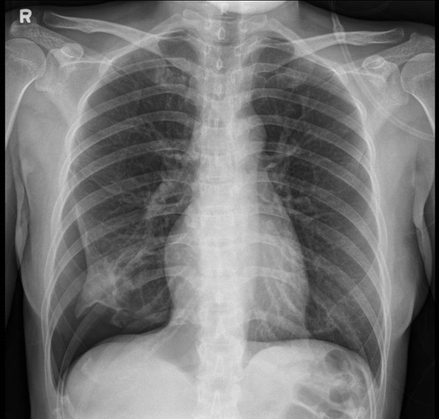
The right pneumothorax previously seen is relatively larger compared to the previous day.
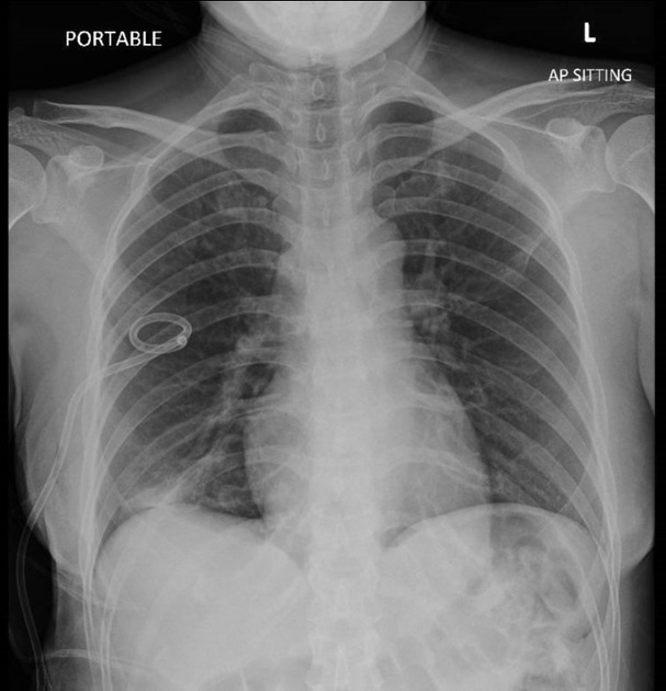
There is interval insertion of a right-sided intercostal catheter. Interval reduction of right pneumothorax is noted with maximal pleural separation of about 1.2 cm in the current study. No frank consolidation or sizeable pleural effusion is seen.
Case Discussion
Despite pneumothoraces often being observed either apically or as a rim around the upper portion of the lung edge, this case depicts a pneumothorax at the basal aspect of the right lung.
Conservative management was first attempted but proved unsuccessful, as seen on the chest image done 20 hours after initial presentation. An intercostal catheter was subsequently inserted, which then allowed for reduction of the pneumothorax.
Spontaneous pneumothoraces are a medical emergency. In some parts of the world (i.e. Australia 1), it is required that the radiographers alerts the referring doctor on this finding so that early intervention can proceed.


 Unable to process the form. Check for errors and try again.