
Presentation
Incidental findings.
Patient Data
Age: 50 years
Gender: Female
From the case:
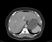
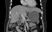
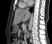
Beaver tail liver

Download
Info

There is a beaver tail lobe of the liver parenchyma (normal variant best seen on axial images) along with diffuse fatty changes of the liver parenchyma (due to relatively low density on venous phase as compared to splenic parenchyma) and hepatomegaly.
Case Discussion
The patient was a known case of thyroid cancer and CT was done for metastatic workup.
Incidental findings were made of beaver tail liver in which the left lobe of the liver is extending laterally and in close contact with the splenic parenchyma (even surrounding it).


 Unable to process the form. Check for errors and try again.