
Presentation
Left neck mass.
Patient Data


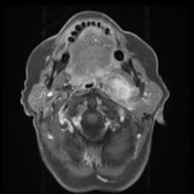

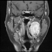

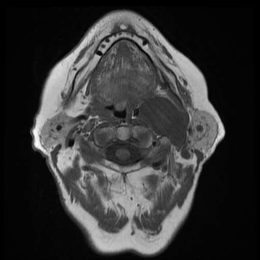
The previously described left carotid space lesion, with imaging features consistent with a benign nerve sheath tumour, is again demonstrated. There is evidence of gradual growth when compared to imaging dating back three years ago. Otherwise it is unaltered in appearance, again demonstrating prominent high T2 signal peripherally and central low T2 signal, a feature which favours a neurofibroma/schwannoma. It remains well-circumscribed, no evidence of direct invasion, and no lymph node enlargement.
The internal carotid artery is displaced medially, and there is narrowing of the oropharynx. The internal jugular is displaced postero-laterally, compressed between the mass and the deep lobe of the parotid.
Conclusion: Continued gradual enlargement of the left carotid space benign nerve sheath tumour.
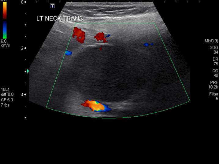
Ultrasound guided biopsy.
Using ultrasound guidance and sterile technique the skin was infiltrated near the angle of the mandible, and an 18 gauge core biopsy needle passed into the carotid sheath mass.
Manual deployment of a 10 mm core biopsy was performed, carefully avoiding arterial structures nearby.
Single sample obtained appeared approximately 4 mm long, and this was sent in formalin for histology.
The patient was recovered in the department, and no immediate complication noted.
Case Discussion
The MR appearances are those of a mass in the carotid space, most likely a nerve sheath tumour.
Ultrasound biopsy showed features of benign peripheral nerve sheath tumour: the morphology is more suggestive of neurofibroma than schwannoma.
HISTOPATHOLOGY
CLINICAL NOTES: Left carotid sheath mass ?Schwannoma ?Pleomorphic adenoma ?Carotid body tumour.
MICROSCOPIC DESCRIPTION: The core biopsy shows a proliferation of spindled stromal cells. They have elongated nuclei with no significant nuclear pleomorphism. The cytoplasm is ill-defined. No mitoses or necrosis is seen. The background is loose and myxomatous with thin wavy bundles of collagen. The spindled cells are S-100 positive. They are synaptophysin, smooth muscle actin and CAM5.2 negative. The features are those of benign peripheral nerve sheath tumour. The morphology is more suggestive of neurofibroma than schwannoma.
DIAGNOSIS: Left parapharyngeal mass: Benign peripheral nerve sheath tumour.


 Unable to process the form. Check for errors and try again.