
Bilateral middle cerebellar peduncle lesions secondray to acquired hepatocerebral degeneration
Presentation
Disturbed conscious level.
Patient Data


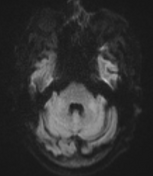
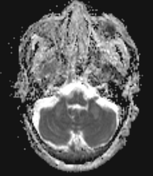
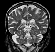
Bilateral middle cerebellar peduncles abnormal signal eliciting high signal on T2 & FLAIR WI and low signal on T1 WI with no diffusion restriction.
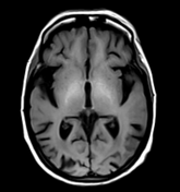
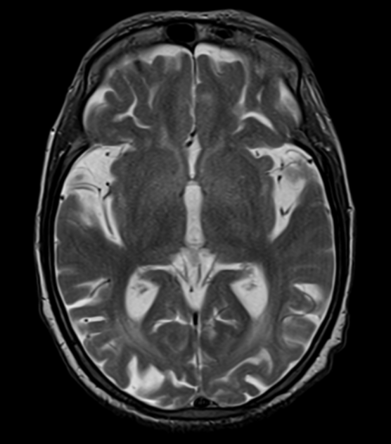
Bilateral hyperintensity in the globi pallidi.
No recent vascular insult.
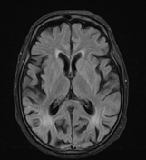
Cerebral arterioleuckoencephalopathy and bilateral cerebral chornic ischaemic foci.
Brain atrophic changes.
Case Discussion
The patient was known hepatitis C virus positive patient with cirrhotic chronic liver disease and multiple prior episodes of hepatic encephalopathy, presented currently to ER with disturbed conscious level. MRI showed no evidence of recent vascular insult, yet it showed signs of acquired hepatocellular degeneration including bilateral basal ganglia T1 hyperintensity of both globi pallidi and bilateral middle cerebellar peduncles lesions of abnormal signal eliciting low signal on T1, high signal on T2 and FLAIR WI with no diffusion restriction.
Acquired hepatocerebral degeneration is an uncommon irreversible extrapyramidal neurodegenerative condition encountered in patients with cirrhotic chronic liver disease, resulting in widespread cerebral, basal ganglia, and cerebellar damage.
Typical appearnce on MRI includes:
T1 intrinsic hyperintensity in the globus pallidus, subthalamic region, and midbrain (substantia nigra) which is thought to be a reflection of increased tissue concentrations of manganese
increased signal in the middle cerebellar peduncles on T2 & FLAIR WI
Case courtesy of Prof. Ayda Youssef.MD of radiodiagnosis. National cancer institute. Egypt


 Unable to process the form. Check for errors and try again.