
Presentation
Left-sided facial numbness and headache.
Patient Data
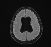

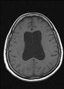
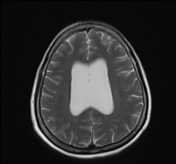
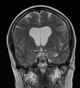
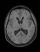
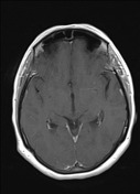
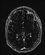
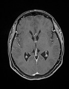
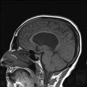
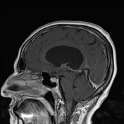
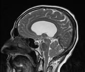
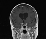
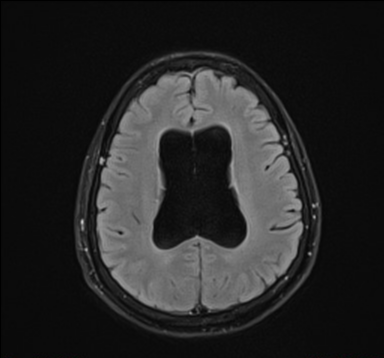
symmetrical dilatation of the lateral ventricles with normal-sized third and fourth ventricles and the rest of cerebrospinal fluid spaces, and with no abnormal periventricular or subependymal signal intensity, or detected focal lesion at the area of interventricular foramina
the septum pellucidum appears thinned and fenestrated
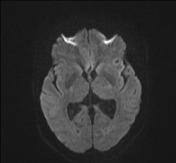
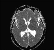
a hyperintense focus in the left periventricular frontal white matter on T2W/FLAIR images without mass effect, restricted diffusion, or contrast enhancement
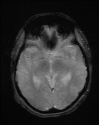
the rest of the study shows normal other findings including both trigeminal nerves along their visualized tracts and both cerebellopontine angles
Case Discussion
Isolated constant inactive hydrocephalus of the lateral ventricles with thinning and fenestration of the septum pellucidum (representing chronicity) and without detected focal lesion, raising the possibility of bilateral stenosis of the interventricular foramen (of Monro).
The mentioned left frontal hyperintense focus characteristics are suggesting a non-specific incidental one, needing clinical correlation and follow-up accordingly.
Bilateral stenosis of the interventricular foramen (of Monro) is a rare condition.
Radiological diagnosis is done by bilateral lateral ventricular dilatation and slit-like third ventricle, with the exclusion of other possible causes.
Possible underlying causes include infection (particularly TORCH infections) causing inflammation and subsequent scarring, congenital atresia or membranes, vascular malformations, and tumors, in the area of the interventricular foramen.
MRI with i.v. contrast agent administration and high-resolution three-dimensional sequences is the technique of choice. It is also useful in guiding the endoscopic treatment when indicated.


 Unable to process the form. Check for errors and try again.