
Presentation
Chronic headache, tinnitus, sensorineural hearing loss on the left side, and left facial neuropathy.
Patient Data
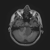

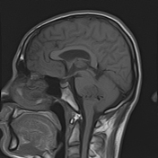

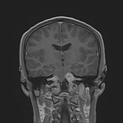
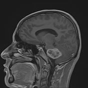
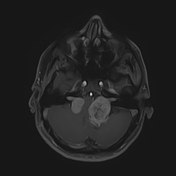
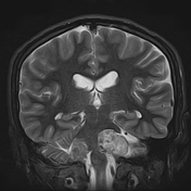
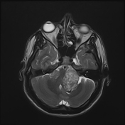
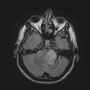
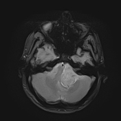
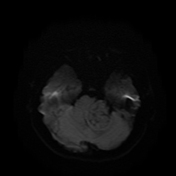
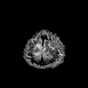
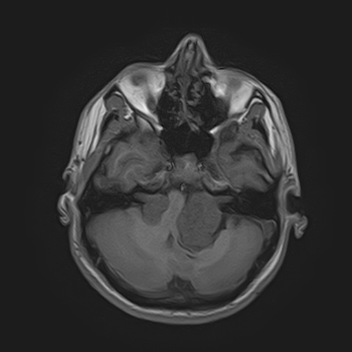
bilateral lesions arising from the internal acoustic meatus and protruding into the cerebellopontine angles deforming the brain stem, the 4th ventricle, and the cerebral aqueduct (Koos grade IV), they are hypointense on T1, heterogeneous on T2 and FLAIR (predominantly hyperintense), avidly enhancing on T1C+, and they show increased diffusion on DWI/ADC map
no blooming artifact is noted on T2* sequence
no hydrocephalus is noted
no other abnormality is noted
Pathology
Sections show biphasic neoplastic growth with some areas that have loose background and consists mostly of stellate shaped cells, while other areas have more dense background and consists of spindle cells arranged in short bundles with nuclear palisading and formation of several Verocay bodies. There is no evidence of malignant changes.
Final diagnosis
Schwannoma (acoustic neuroma), WHO grade I.
Case Discussion
The patient underwent surgery for the left CPA tumor (total microsurgical resection via retro-sigmoid approach), the patient did well post-op, and the pathology report confirmed the diagnosis of schwannoma.
The presence of bilateral vestibular schwannoma on MRI is the hallmark of neurofibromatosis type 2 and confirms the diagnosis 1.
This case represents NF2 (Gardner subtype), which is characterized by late onset, and low incidence of associated other intracranial tumors 2.
Courtesy: Dr Tamer Alameer (Neurosurgery Department, Al Assad University Hospital, Damascus)


 Unable to process the form. Check for errors and try again.