
Presentation
Abdominal tenderness and pain, longstanding. Occasional shortness of breath.
Patient Data


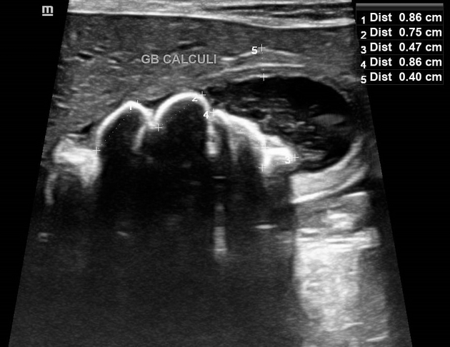
Macromillimeter sized multiple hyperechoic, posteriorly shadowing foci that subtly twinkles on color Doppler mapping are noted within the gallbladder lumen, conglomerating at the neck region and surrounded peripherally with low level echo fluid. Thickened gallbladder inner and outer wall coats is noted. No biliary/ pancreatic ducts dilatation.
There are also:
mild hepatic veins engorgement coupled with mild pericardial effusion
moderately reflective bilateral renal cortical parenchymal echotexture
minimal perinephric/sub-capsular clear fluid
mild abdominal ascites
mild left sided pleural effusion
Case Discussion
Sonograms of the abdomen shows gallbladder cholelithiasis (measuring up to 9 mm in diameters) with features of cholecystitis and mild biliary sludge albeit with no obstructive tendency.
The rest of the anomalies identified including: Mild bilateral perinephric/sub-capsular fluids (urinoma?) with features of bilateral parenchymal renal disease; Mild hepatic veins engorgement with mild pericardial effusion and; Minimal abdominal ascites plus minimal left sided pleural effusion seen, are purely incidentals which required follow ups and were concerning for cardiorenal syndrome.


 Unable to process the form. Check for errors and try again.