Biliary tract embryonal rhabdomyosarcoma
Updates to Case Attributes
The most common cause of benign obstructive jaundice in childhood arecholedochal cysts, while the most common malignant cause of obstructive jaundice in childhood is rhabdomyosarcoma. However in this case, the other differential diagnosis is lymphoma (homogeneous mass enhancement and associated with lymphadenopathy), while rhabdomyosarcoma mainly shows heterogeneous enhancement.
-<p>The most common cause of benign obstructive jaundice in childhood are<strong> </strong>choledochal cysts, while the most common malignant cause of obstructive jaundice in childhood is rhabdomyosarcoma. However in this case, the other differential diagnosis is lymphoma (homogeneous mass enhancement and associated with lymphadenopathy), while rhabdomyosarcoma mainly shows heterogeneous enhancement.</p>- +<p>The most common cause of benign obstructive jaundice in childhood are<strong> </strong><a title="Choledochal cysts" href="/articles/choledochal-cysts">choledochal cysts</a>, while the most common malignant cause of obstructive jaundice in childhood is <a title="Rhabdomyosarcomas (biliary tract)" href="/articles/rhabdomyosarcomas-biliary-tract">rhabdomyosarcoma</a>. However in this case, the other differential diagnosis is lymphoma (homogeneous mass enhancement and associated with lymphadenopathy), while rhabdomyosarcoma mainly shows heterogeneous enhancement.</p>
Updates to Study Attributes
There is a mixed echogenicity ill-defined mass lesion located in the portahepatis measuredporta hepatis measuring 5 x 4 cm, it shows increased blood flow on color Doppler mode. The mass encasingencases the hepatic artery, portal vein and extra hepaticextrahepatic biliary tree, causing intra hepaticintrahepatic dilatation of the biliary tree. Multiple enlarged portahepatisporta hepatis lymph nodes.
Updates to Study Attributes
TISSUE ORIGINTissue origin: 1- Omentum. omentum. 2- Porta. porta hepatis lymph node. 3- Porta. porta hepatis mass.
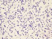
DIAGNOSISDiagnosis: Omentum, porta hepatis lymph node and porta hepatis mass; biopsies: Consistent with embryonal rhabdomyosarcoma. The tumor cells are positive for Desmindesmin, Myogenin (Focalmyogenin (focal) and Glypicanglypican-3. They are negative for Panpan-CK, SALL4 and Heppar1heppar1 immunostains.
Image Pathology (Desmin) ( update )
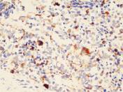
Image Pathology (Myogenin) ( update )

Image Pathology (Glypican-3) ( update )
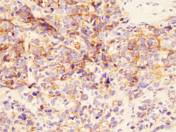
Image Pathology (Pan-CK, SALL4 & Heppar1) ( update )