
Presentation
Dementia
Patient Data
Age: 75 years
Gender: Female

Download
Info
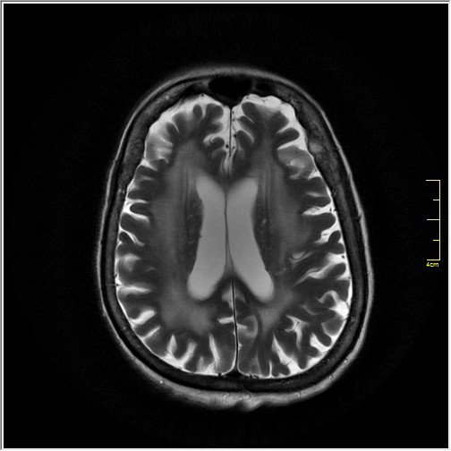
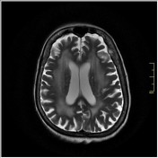
There are marked confluent symmetrical T2 hyperintensities in bilateral periventricular and bilateral centrum semiovale with sparing of subcortical U fibers, representing chronic ischemic changes. Some small foci of blooming are noted on gradient sequence.
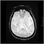
A small old hematoma is noted in the left thalamus.
Chronic lacunar infarcts are noted in the bilateral gangliocapsular regions.
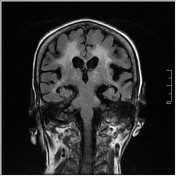
Generalized age-related corticocerebral atrophy is seen.


 Unable to process the form. Check for errors and try again.