
Presentation
Anterior knee pain with a history of minor trauma.
Patient Data

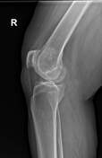
lucent line is seen at the superolateral aspect separating the right patella into two fragments
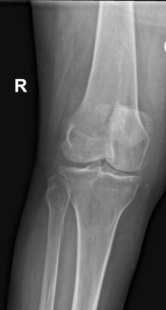
non-uniform, asymmetric joint space narrowing more evident at the medial compartment with osteophytic formation at the medial and lateral tibial plateaus and both femoral condyles, in keeping with degenerative joint diseases (osteoarthritis)
patch of sclerosis with internal lucencies present in the distal femoral metaphysis, no endosteal scalloping, periosteal reaction, or extra-osseous mass, with a narrow zone of transition
Case Discussion
The location of a patellar bone fragment at the superolateral aspect of the patella, and a lack of joint effusion may suggest bipartite patella and help to confirm that this is not a fracture, however, traumatic separation of a bipartite patella is also possible and therefore comparison with previous radiographs may be helpful to check the position of the parts of the patella.
The lower femoral metaphyseal sclerotic area is suggestive of an intramedullary lesion most likely a medullary bone infarct.


 Unable to process the form. Check for errors and try again.