
Presentation
Lower abdominal dull pain with recurrent urinary tract infection for a few months.
Patient Data
Age: 30 years
Gender: Male
From the case:
Bladder calculus


Show annotations
Download
Info
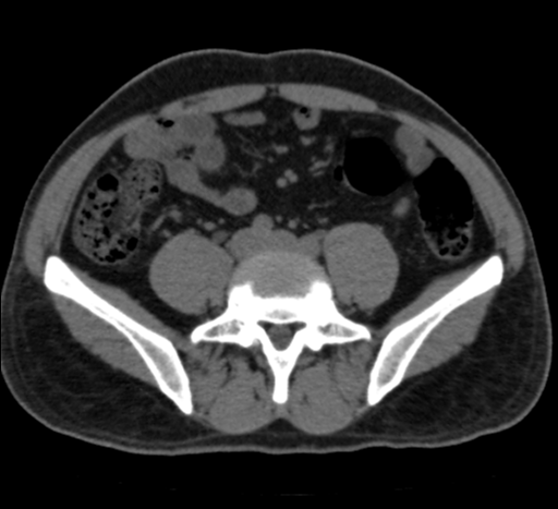
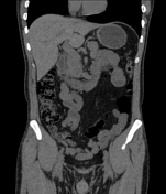
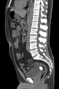
An oval-shaped, radiodense calculus is seen in the dependent part of the urinary bladder, however wall thickness is within normal limits.
Another small radiodense calculus is seen in the distal-most, right-sided ureter, however, no definite hydro-ureter or hydronephrosis is seen.
The rest of the imaged viscera appear unremarkable.
Case Discussion
CT features are of vesicle calculus along with right-sided ureteric calculus as described above.
Co-contributor: Dr. Anwar-ul-Haq Zadran.


 Unable to process the form. Check for errors and try again.