
Presentation
Acute onset left side weakness.
Patient Data
Age: 45 years
Gender: Male


Download
Info
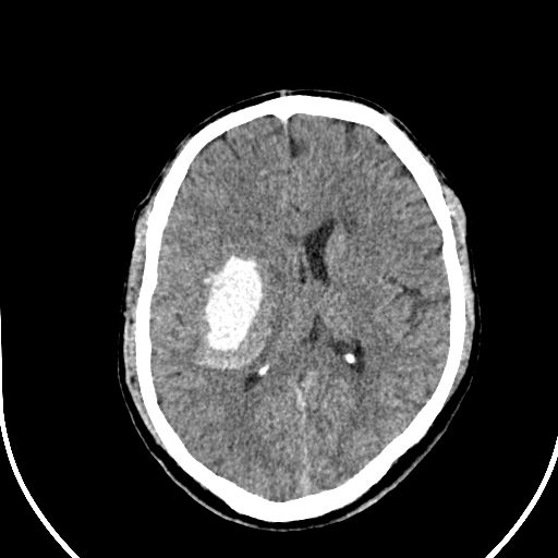
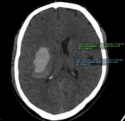
Non-contrast CT head demonstrating 'blend sign' within right intracerebral haemorrhage.
Download
Info
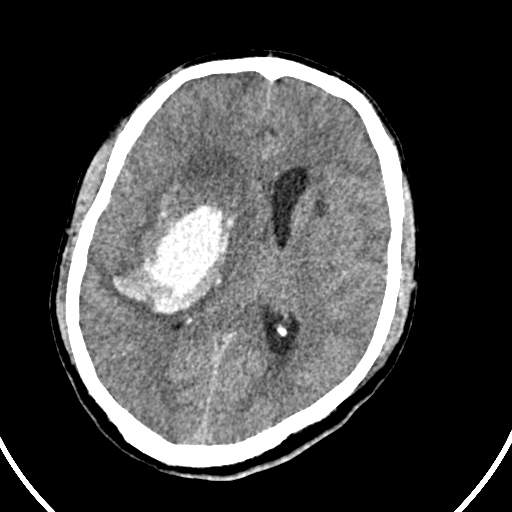
Non-contrast CT head 24 hours later demonstrating increased volume of haematoma with worsening features of mass effect.
Case Discussion
This case demonstrates the blend sign of intracranial haemorrhage. This blend sign is a useful predictor for haematoma growth following intracerebral haemorrhage, as is demonstrated in this case.
The blend sign is defined as 1:
intracranial haematoma with two different density components identifiable by the naked eye
difference between the two densities >18 HU
the hypoattenuating component is not encapsulated by the hyperattenuating region


 Unable to process the form. Check for errors and try again.