
Presentation
Vomiting and ataxia.
Patient Data
Age: 5 years
Gender: Male
From the case:
Brainstem glioma

Download
Info

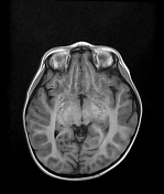
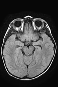
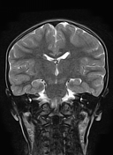
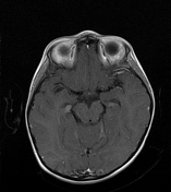
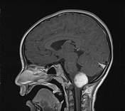
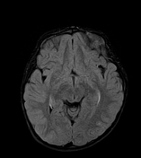
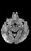
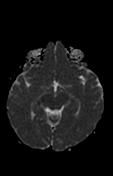
The MRI sequences demonstrate a well-circumscribed dorsally exophytic brainstem mass centered on the medulla oblongata of low signal on T1, high signal on T2/FLAIR with intense and relatively homogeneous enhancement on postcontrast sequences. Partial effacement of the median aperture of Magendie and cerebellomedullary cistern with mild dilatation of the 4th ventricle. No significant dilatation of the supratentorial ventricular system.
Case Discussion
MRI features are consistent with a brainstem glioma.


 Unable to process the form. Check for errors and try again.