
Presentation
A known case of right breast cancer was referred for a liver MRI due to the presence of liver masses on the US exam.
Patient Data
Age: 55 years
Gender: Female
From the case:
Breast carcinoma with liver metastasis


Show annotations
Download
Info
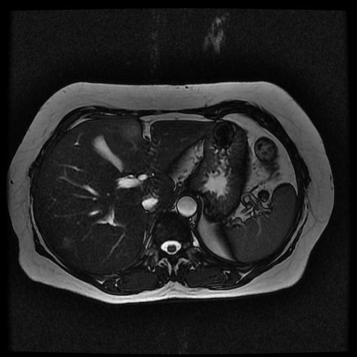
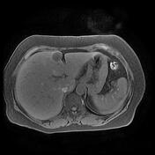
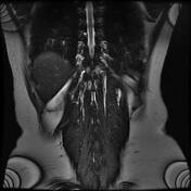
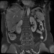
Axial T2-weighted imaging shows three abnormal signal liver masses along with segments IVb, VIII, and VII displaying a target sign characterized as a hyperintense center marginated by a lesser intense rim of viable tumor.
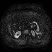
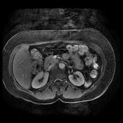
Liver masses showed marked water restriction on DWI images and post-contrast peripheral rim of enhancements.
Case Discussion
Known case of right breast cancer; pathology-proven invasive ductal carcinoma with hepatic metastases.


 Unable to process the form. Check for errors and try again.