
Presentation
Haemoptysis.
Patient Data
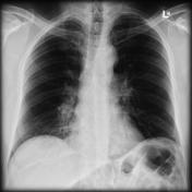
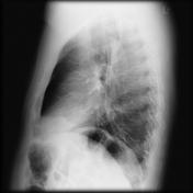

The right hilum is bulky, especially inferiorly with the normal pulmonary vasculature poorly seen. Minor patchy opacities is seen in the medial basal aspect of the right lung.

The right hilum demonstrates a rounded mass with heterogeneous attenuation closely related to the right lower lobe bronchus. Right sided infrahilar airspace infiltrates are present, with extension to the pleura in some areas, representing areas of pulmonary haemorrhage.
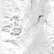

A microcatheter has been inserted into the right bronchial artery. Contrast injection demonstrates a tortuous and dilated artery supplying the right lower lobe. After extensive discussion with the thoracic surgery team, the decision to perform a bronchial angiogram and embolisation.
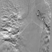
The second image demonstrates placement of two coils in the proximal segment of the right bronchial artery, with no distal flow. The artery has been successfully embolised.
Case Discussion
Known Behcet's disease.


 Unable to process the form. Check for errors and try again.