
Presentation
Trauma two months back with knee joint pain.
Patient Data


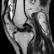
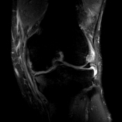
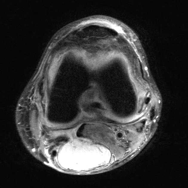
Absent bow-tie sign with complete non-visualization of the body of the medial meniscus. The fragment appears to be displaced centrally in the intercondylar notch with a double PCL sign suggesting a bucket handle tear.
Grade I signal in the anterior horn of the lateral meniscus.
Fiber discontinuity of the anterior cruciate ligament (ACL) with surrounding fluid, suggesting a complete tear, and mild buckling of the posterior cruciate ligament (PCL).
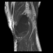
Moderate knee joint effusion with suprapatellar extension.
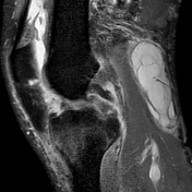
A large cystic lesion with multiple internal thin septations with a narrow neck between the medial gastrocnemius and semimembranous tendon, suggesting a Baker‘s cyst.
Mild marrow edema over the medial tibial condyle.
Mild thinning of the femoral, tibial and patellar articular cartilage with few marginal osteophytes, with a few tiny subchondral cysts over the proximal tibia and patella - osteoarthritic changes.
Case Discussion
Typical bucket handle meniscal tear case with classic signs as follows:
meniscal fragment in the intercondylar notch


 Unable to process the form. Check for errors and try again.