
Presentation
No history of trauma. Right shoulder pain worsened by exertion.
Patient Data
Age: 25 years
Gender: Female
From the case:
Buford complex

Download
Info

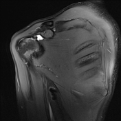
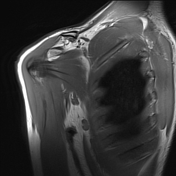
- Moderate fluid buildup in the subscapular recess (best seen on PD weighted sequences), which is not necessarily pathological.
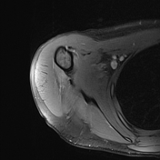
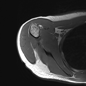
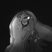
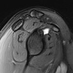
- Absent anterosuperior labrum in the 12-3 o'clock position, with resultant thickening of the middle glenohumeral ligament, best appreciated on the axial PD FS sequence (see key image).
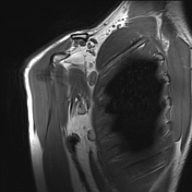
- Axillary lymph nodes showing reactive morphology.
From the case:
Buford complex

Download
Info
- Saggital PD FS: key image demonstrating fluid buildup in the subscapular recess.
- Axial PD FS: short arrow demonstrating absence of anterosuperior labrum, long arrow indicating the thickened MGHL.
Case Discussion
The incidentally demonstrated anatomical labral variation represents the Buford complex.


 Unable to process the form. Check for errors and try again.